Ürün Açıklaması
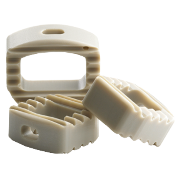
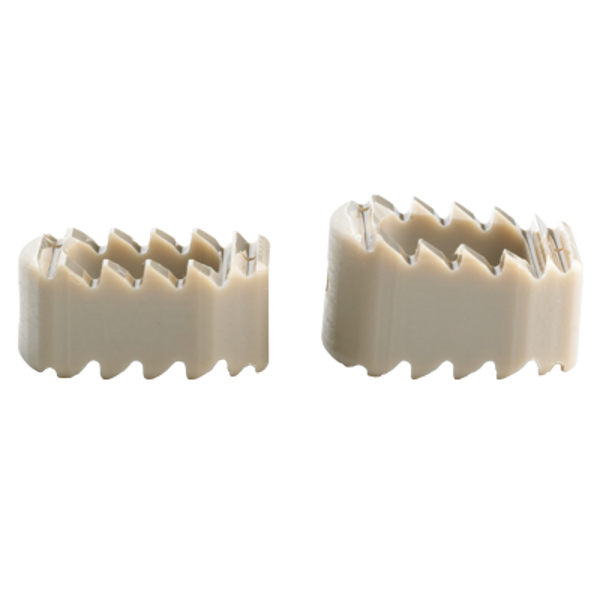
Vista -S Servikal füzyon Cihaz üretilmiştir PEEK-OPTIMA *, gücünde bir yük paylaşımı, radyolusent, biyouyumlu malzeme ve istikrar. Üç ayak izleri ve yükseklikleri bir dizi sunulan Vista -S implantlar hastalarınızın değişen anatomisi konaklayabilir.Eşsiz köpekbalığı diş yüzeyinin deseni göç riskini azaltır ve lider konik kenar olur yerleştirilmesini kolaylaştırabilir.
Özellikler
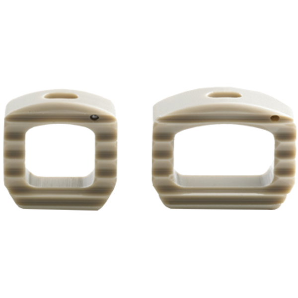
- Radyolusent maddeden ve tantal boncuk belirteçler füzyon ve implant yerleştirme görselleştirme için izin
- Konik arka kenarı geliştirilmiş görünürlük ve yerleştirme kolaylığı için merkezi yerleştirici ile kombine
- Benzersiz “köpekbalığı dişi” yüzey deseni göç riskini azaltır
- Greft yerleştirilmesi için büyük bir pencere aç geometri
- Farklı hasta anatomisi karşılamak için çeşitli boyutları mevcuttur
* PEEK-OPTIMA ® Polimer Invibio Ltd’nin tescilli bir ticari markasıdır
ZS-SA0700-26_A
Device Description
The Vista®-S Device is manufactured wholly from unfilled PEEK-OPTIMA® LT1, a polyetheretherketone. This material is a thermoplastic polycondensate, semicrystaline polymer. It is used in this device in the unfilled state (i.e. no glass or carbon fiber fill). Due to the radiolucent nature of PEEK-OPTIMA® LT1, three radiopaque markers made of tantalum are incorporated into the device to indicate the nose end and the superior and inferior corners of the opposite end for use in postoperative monitoring of device position.
The superior and inferior surfaces of the device have a textured surface to provide increased stability. The device is available in a variety of cross-sectional geometries and sizes. These implants offer two different included angle options to maintain the natural contour of the spine.
These implants are intended for single use only and must not be reused under any circumstances. Surgical instruments are also available to assist in the implantation of the device.
Indications
The Vista-S Device is a cervical interbody fusion device indicated for use in skeletally mature patients with degenerative disc disease (DDD) with/without radicular symptoms at one level from C2-T1. DDD is defined as discogenic pain with degeneration of the disc confirmed by history and radiographic studies. These patients should have had six weeks of non-operative treatment. The Vista-S Device is intended for use with supplemental fixation systems and with autogenous bone graft. The Vista-S Device is implanted via an anterior approach.
Contraindications
- Active local infection in or near the operative region.
- Active systemic infection and/or disease.
- Severe osteoporosis or insufficient bone density, which in the medical opinion of the physician precludes surgery or contraindicates instrumentation.
- Prior surgical procedure using the desired operative approach.
- Spinal conditions other than cervical DDD.
- Current metastatic tumors of the vertebrae adjacent to the implant.
- Known or suspected sensitivity to the implant materials.
- Endocrine or metabolic disorders known to affect osteogenesis (e.g., Paget's disease, renal osteodystrophy, hypothyroidism).
- Systemic disease that requires the chronic administration of nonsteroidal anti-inflammatory or steroidal drugs.
- Significant mental disorder or condition that could compromise the patient's ability to remember and comply with preoperative and postoperative instructions (e.g., current treatment for a psychiatric/psychosocial disorder, senile dementia, Alzheimer's disease, traumatic head injury).
- Neuromuscular disorder that would engender unacceptable risk of instability, implant fixation failure, or complications in postoperative care. Neuromuscular disorders include spina bifida, cerebral palsy, and multiple sclerosis.
- Pregnancy.
- Patients unwilling to follow postoperative instructions, especially those in athletic and occupational activities.
- Morbid obesity.
- Symptomatic cardiac disease.
- Skeletal immaturity.
- Grossly distorted anatomy.
- Conditions other than those indicated.
Warnings
- Surgery is not always successful. Preoperative symptoms may not be relieved or may worsen. Surgical knowledge of the procedure and the device are important, as is patient selection. Patient compliance is also important. Tobacco and alcohol abuse may lead to unsuccessful results.
- Appropriate device selection is crucial to obtain proper fit and to decrease the stress placed on the implant.
- Components of competitive spinal systems should not be used with the Vista-S Fusion Device.
- Delayed healing can lead to fracture or breakage of the implants due to increased stress and material fatigue. Patients must be fully informed of all the risks associated with the implant and the importance of following postoperative instructions regarding weight bearing and activity levels to facilitate proper bone growth and healing.
- The implant must be handled carefully following manufacturer’s instructions to prevent damage to the implant.
- Implants must not be modified or otherwise processed in any way.
- Once a device has been implanted, it must never be reused. If the package is damaged, opened, or if the expiration date has passed, but the device is not used, the device must be returned to Zimmer. The device must not be resterilized by the end user.
- Results may be worse with multilevel disease. Supplemental fixation is required. The surgeon must be familiar with fixation techniques and appropriate hardware.
- The surgeon must be familiar with the appropriate technique to implant the supplemental internal fixation and the appropriate hardware.
- MRI Compatibility 10.1 The patient must be told that implants can affect the results of computer tomography (CT) or magnetic resonance imaging (MRI) scans. 10.2 The Vista-S Fusion Device has not been evaluated for safety or compatibility in the MR environment. 10.3 The Vista-S Fusion Device has not been tested for heating or migration in the MR environment.
Surgeon Precautions
- The implantation of an intervertebral body fusion device should be performed only by experienced spinal surgeons with specific training in the use of this device because this is a technically demanding procedure presenting a risk of serious injury to the patient.
- The surgeon must have a thorough knowledge of the mechanical and material limitations of semicrystaline polymeric surgical implants and be thoroughly familiar with the surgical technique for implanting the Vista®-S Device for the given Indications for Use.
- Based on fatigue testing results, the physician/surgeon should consider the levels of implantation, patient weight, patient activity level, other patient conditions, etc. which may impact on the performance of the system.
- The surgeon should be familiar with the various devices and instruments and verify that all are available before beginning the surgery. Additionally, the packaging and implant should be inspected for damage prior to implantation.
- In the event that removal of the implant is considered (e.g. due to loosening, fracture, migration of the implant; infection; increased pain, etc.), the risks versus benefits must be carefully weighed. Such events can occur even after healing, especially in more active patients. Appropriate postoperative care must be given following implant removal to avoid further complication.
- The surgeon must be thoroughly familiar with the options for supplemental internal fixation systems and the associated surgical techniques.
- Implants must be fully seated within the inserter prior to use. Care must be taken not to over-tighten the implant-inserter assembly. Additionally, care must be taken not to manipulate the inserter implant interface in a way not recommended by the surgical technique.
- The surgeon must ensure the implant is properly seated prior to closing of the soft tissue.
- Extreme caution must be used around the spinal cord, nerve roots and blood vessels.
Patient Precautions
- Postoperative care instructions are extremely important and must be followed carefully. Non-compliance with postoperative care instructions could lead to failure of the device, and the possibility of additional surgery to remove the device.
- The patient should limit activities that result in overhead lifting, repetitive neck bending (especially neck extension) and heavy lifting until a physician determines solid bony fusion is achieved.
- An orthotic brace may be worn following surgery for support. The attending physician, based upon each patient’s clinical progress, will determine whether a brace is appropriate and, if necessary, the length of time the brace is prescribed.
- Non-steroidal anti-inflammatory and steroidal drugs should be avoided for at least 45 days, or as directed by a physician, postoperatively.
Possible Adverse Effects
As with any surgical procedure, certain complications may result.
- Potential complications associated with the device itself include:
- Bone lysis associated with particulate debris,
- Device bending,
- Device fracture,
- Device loosening,
- Device migration.
- Failure to properly fill and compress the graft material into the area surrounding the implant may result in delayed healing and or nonunion.
- If either the iliac crest, fibula or rib are utilized as a secondary donor site for bone graft material, associated complications which may occur include; hematoma requiring treatment, persistent donor site pain, pelvic instability (iliac crest only), nerve damage with sensory loss, deep or superficial wound infection, herniation (iliac crest only) or excessive bleeding.
- Possible other Adverse Effects associated with use of the Vista®-S Device include:
- Abscess,
- Adjacent segment disease,
- Adverse reaction to anesthesia,
- Airway obstruction,
- Allergic reactions to prophylactic antibiotics or blood transfusions,
- Allergic reaction to the implant(s),
- Anesthetic or post-anesthetic reactions,
- Anterior longitudinal ligament penetration,
- Arachnoiditis,
- Arrhythmia,
- Atelectasis,
- Back pain,
- Bladder dysfunction,
- Bone or vertebral fracture during insertion of the device,
- Bone lysis associated with particulate metal debris from the supplemental internal fixation,
- Bone resorption,
- Bursitis,
- Cauda equina syndrome,
- Cellulitis,
- Cerebrovascular accident,
- Constipation,
- Cord injury,
- Death,
- Decreased leg strength,
- Decreased reflexes,
- Deep vein thrombosis,
- Delayed union or nonunion,
- Disc herniation,
- Donor site events (if additional donor site is necessary),
- Dural tear or leak,
- Dysphasia,
- Dysphonia,
- Failure of instrumentation,
- Foot drop,
- Fracture of pedicle bone,
- Graft failure (fracture, resorption, etc.),
- Graft expulsion,
- Great vessel damage,
- Hematoma,
- Hemorrhage,
- Ileus,
- Incisional hernia,
- Incisional pain,
- Infection throughout the body (systemic),
- Infection of the wound,
- Ischemia,
- Leg pain,
- Loss of reduction,
- Loss of spine curvature,
- Malpositioned screws,
- Myelopathy,
- Myocardial infarction,
- Neurapraxia,
- Nerve root injury,
- Organ, nerve, blood vessel or muscle damage,
- Osteoporosis local to implant site,
- Pain,
- Painful hardware,
- Paralysis,
- Perforation of the trachea, esophagus or pharynx,
- Pneumonia,
- Pseudarthrosis,
- Pulmonary embolism,
- Radiculopathy or lack of improvement in existing radiculopathy,
- Recurrent deformity,
- Reflex sympathetic dystrophy (RSD),
- Scar formation,
- Screw loosening,
- Screw migration,
- Seroma,
- Spinal cord injury,
- Spinal stenosis,
- Spondylolisthesis,
- Subsidence of the implant,
- Swelling,
- Syringomyelia,
- Thromboembolism,
- Thrombophlebitis,
- Thrombosis,
- Tumor formation and/or recurrence,
- Urinary tract infection,
- Vocal cord paralysis,
- Wound dehiscence.
- Complications which may be associated with thoracotomy include:
- Acute respiratory distress syndrome,
- Atelectasis,
- Pneumothorax,
- Pulmonary contusion,
- Upper respiratory tract infection.
- Additional complications that are not anticipated may also occur.
- Reoperation may be necessary to correct adverse effects.